Ombro: cíngulo escapular, úmero e manguito rotador
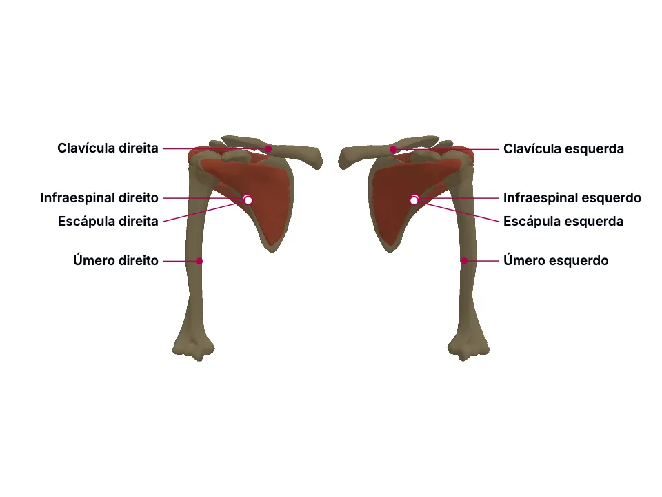
O manguito rotador - subescapular, infraespinal, redondo menor e supraespinal (SITS) - centraliza uma cabeça umeral grande demais para a cavidade glenoide RASA da escápula, trocando estabilidade óssea por mobilidade: por isso o ombro é a articulação mais luxada do corpo (direção anteroinferior em mais de 95% dos casos), o que arrisca o nervo axilar no espaço quadrangular (borda pelo redondo menor), testável pela sensibilidade do 'distintivo regimental'. O supraespinal, que inicia a abdução, atravessa um espaço subacromial estreito e está entre os tendões mais comumente lesados do manguito, com fraqueza testável pelo teste de Jobe. A clavícula, elo do cíngulo peitoral com a escápula, está entre os ossos mais comumente fraturados do corpo, tipicamente no terço médio - seu segmento mais fino e sem reforço ligamentar.
Material educacional escrito com apoio de IA e conferido por revisão automatizada, sem revisão médica nomeada. Não substitui o livro-texto nem orientação clínica.
Essencial
A cavidade glenoide - parte da própria malha da escápula, não uma peça à parte - é rasa: 'The glenoid cavity is a shallow osseous structure deepened by a fibrocartilaginous rim, the glenoid labrum, which spans the vault's osseous periphery.' O tamanho da cabeça umeral em relação a essa cavidade rasa é desproporcional: 'The humeral head's disproportionate size relative to the shallow glenoid fossa (a 4:1 surface area ratio) contribute to the shoulder joint's high mobility and susceptibility to dislocation.'
Quem compensa essa falta de encaixe ósseo são os quatro músculos do manguito rotador - subescapular, infraespinal, redondo menor e supraespinal, o mnemônico SITS: 'The rotator cuff includes the following muscles: Subscapularis Infraspinatus Teres minor Supraspinatus A helpful mnemonic for recalling these muscles is "SITS."' Eles se originam cada um de uma região distinta da MESMA escápula e convergem sobre o úmero: 'The primary biomechanical role of the rotator cuff is to stabilize the glenohumeral joint through compression of the humeral head against the glenoid. The 4 rotator cuff muscles originate from the scapula and insert on the humerus.' Coordenados, eles mantêm a cabeça umeral centralizada durante o movimento: 'Coordinated contraction of the rotator cuff muscles maintains humeral head centering within the glenoid during shoulder motion, permitting a broad range of motion while preserving joint stability.' - e é por isso que a fonte os descreve como 'The rotator cuff muscles thus act as the glenohumeral joint's dynamic stabilizers.' Mas essa cobertura muscular não é completa: 'leaving the inferior aspect relatively unsupported. This anatomic configuration increases the risk of inferior glenohumeral dislocation.'
Dos quatro músculos do manguito, o supraespinal é quem inicia o movimento de abdução: 'The supraspinatus muscle originates in the supraspinous fossa of the scapula and inserts into the greater tubercle of the humerus, facilitating the abduction of the humerus.' e 'The supraspinatus initiates shoulder abduction and is particularly important during the first 0° to 15° of motion, after which the deltoid becomes the principal abductor.'
Para chegar ao tubérculo maior, o tendão do supraespinal atravessa um corredor estreito sob o acrômio: 'The subacromial space is bounded superiorly by the acromion and acromioclavicular joint, anteriorly by the acromion and coracoacromial ligament, and inferiorly by the humeral head. Within this space lie the supraspinatus tendon, portions of the infraspinatus and subscapularis tendons, the long head of the biceps tendon, and the subacromial-subdeltoid bursa.' Esse espaço já é apertado por natureza: 'The subacromial space, typically 1 to 1.5 cm in height, narrows physiologically with abduction and forward flexion.' E o próprio mecanismo que deveria protegê-lo pode piorar o aperto: quando o manguito enfraquece ou se fatiga, a cabeça umeral perde a força que a mantém centralizada e migra para cima, estreitando ainda mais o espaço que o supraespinal precisa atravessar: 'Secondary impingement occurs despite normal static anatomy and results from dynamic factors that allow superior migration of the humeral head during motion, such as rotator cuff weakness or fatigue resulting in loss of centralizing force'
A direção anterior domina as luxações de ombro: 'Anterior dislocation comprises 96% of cases and is often the result of a force directed to the shoulder joint while the arm is abducted and externally rotated.' - confirmado de outra fonte: 'Over 95% of shoulder instability events occur in the anterior direction.' O nervo axilar contorna exatamente a região que essa luxação estica: 'The axillary nerve, formed from the posterior cord of the brachial plexus, wraps around the surgical neck of the humerus, and provides innervation to the deltoid and rotator cuff muscles, specifically the teres minor.' - 'The axillary nerve, which wraps around the humeral surgical neck, can sustain damage during dislocation or reduction attempts.'
Antes de alcançar o deltoide, o nervo axilar passa por um espaço quadrangular estreito, cuja borda superior é o próprio redondo menor: 'The quadrangular space is bordered superiorly by the lower border of teres minor muscle, laterally by the surgical neck of the humerus, inferiorly by the upper boundary of teres major, and medially by the long head of the triceps brachial.' É justamente nesse trajeto - e na cápsula anteroinferior que a luxação distende - que o nervo é mais vulnerável: 'The axillary nerve is most susceptible to injury at the origin from the posterior cord, in the quadrilateral space, the anteroinferior aspect of the shoulder capsule, and within the subfascial surface of the deltoid muscle.' Por isso ele é 'The axillary nerve is the most common damaged nerve in glenohumeral dislocation' e, cirurgicamente, 'The axillary nerve (most common nerve injury) is adjacent to the inferior capsule and closest to the inferior glenoid rim at the 6 o'clock position.'
A clavícula fecha o cíngulo peitoral pela frente: 'The clavicle is a sigmoid-shaped long bone with a convex surface along its medial end when observed from a cephalad position. The clavicle connects the axial and appendicular skeletons and, along with the scapula, forms the pectoral girdle.' E, entre todos os ossos do corpo, 'The clavicle is one of the most commonly fractured bones in the human body.'
A razão é geométrica, não só de exposição: 'Because the midshaft of the clavicle is the thinnest segment and does not contain ligamentous attachments, it is the most easily fractured aspect of the clavicle.' Esse ponto mais fino - sem o reforço ligamentar que protege as extremidades - concentra a maioria das fraturas: 'The middle third of the clavicle is fractured in 69% of cases, the distal third is fractured in 28% of cases, and the proximal third is fractured in 3% of cases.'
Na clínica
Estabilidade dependente de músculo, e não de osso, tem um preço: 'The glenohumeral joint is by far the most frequently dislocated joint of the body, accounting for up to 45% of dislocations.' e, quando ocorre, 'Anterior dislocation comprises 96% of cases and is often the result of a force directed to the shoulder joint while the arm is abducted and externally rotated.'
Não por acaso, 'In adults, rotator cuff injury is the most common tendon injury seen and treated.' A fraqueza resultante é testável à beira do leito: 'The Jobe or empty can test used to evaluate for supraspinatus tendinopathy. The patient elevates the arm to about 90 degrees, and with the thumb down (having an empty can in the patient's hand), isometric force is applied downward.' - descrito de outra fonte como 'With the arms in 90° of abduction in the scapular plane and internally rotated (thumbs down), the patient resists downward pressure. Pain and/or weakness suggest supraspinatus pathology.'
A lesão desse nervo tem sinal sensitivo específico e testável: 'The upper lateral cutaneous nerve provides sensation to the inferior lateral deltoid region, at an area called the regimental badge area.' - achado que integra o quadro descrito como 'Axillary nerve injury may lead to deltoid paralysis and loss of sensation over the lateral shoulder.' No plano motor, é o próprio redondo menor - cuja borda forma o espaço por onde o nervo passa - que ajuda a manter a cabeça umeral na cápsula: 'Teres minor is one of the muscles which stabilize the glenohumeral joint. It acts by laterally rotating the arm and helps to keep the head of the humerus in the shoulder capsule.'
O mecanismo típico reforça o padrão: 'These injuries most often result from a fall onto the lateral shoulder, which transmits force along the length of the bone.'
Abrir o aprofundamento
Aprofundamento
A escápula tem uma concavidade na face ventral, a fossa subescapular: 'The ventral surface of the scapula abuts the thoracic rib cage and has a large concavity called the subscapular fossa where the subscapularis attaches.' Na face dorsal, a espinha da escápula separa duas outras fossas: 'Above the spine is the supraspinous fossa and below it is the infraspinous fossa.' E uma quarta origem fica fora das fossas, na borda lateral: 'A ridge runs down the infraspinous fossa near the lateral border from the glenoid cavity to just above the inferior angle. This ridge separates the teres major and minor muscles from the infraspinatus muscle.'
Subescapular - origina-se na fossa subescapular (face ventral) e se insere no tubérculo menor do úmero: 'The muscle originates from the subscapular fossa and inserts onto the lesser tubercle of the humerus.' É o único rotator interno do grupo SITS: 'The subscapularis is the primary medial (internal) rotator of the shoulder, whereas the infraspinatus and teres minor function primarily as lateral (external) rotators.' e o maior componente da parede posterior da axila, prevenindo a luxação anterior durante a abdução: 'The subscapularis is the largest component of the posterior wall of the axilla. This muscle prevents anterior dislocation of the humerus during abduction and produces medial rotation of the arm.'; Supraespinal - origina-se na fossa supraespinal e se insere no tubérculo maior, iniciando a abdução do ombro: 'The supraspinatus muscle originates in the supraspinous fossa of the scapula and inserts into the greater tubercle of the humerus, facilitating the abduction of the humerus.' - 'The supraspinatus initiates shoulder abduction and is particularly important during the first 0° to 15° of motion, after which the deltoid becomes the principal abductor.'; Infraespinal - origina-se na fossa infraespinal e na espinha da escápula, inserindo-se no tubérculo maior e permitindo rotação externa: 'The infraspinatus muscle originates in the infraspinous fossa and scapula spine and inserts into the greater tubercle of the humerus as well and allows external rotation of the humerus.'; Redondo menor - origina-se na borda lateral da escápula, fora das três fossas, e se insere no tubérculo maior, permitindo rotação externa: 'The teres minor originates on the lateral border of the scapula and inserts into the greater tubercle and allows external rotation.' Sua borda superior fecha o espaço quadrangular por onde passam o nervo axilar e os vasos circunflexos posteriores do úmero: 'The quadrangular space is bordered superiorly by the lower border of teres minor muscle, laterally by the surgical neck of the humerus, inferiorly by the upper boundary of teres major, and medially by the long head of the triceps brachial.' e o músculo ajuda a manter a cabeça umeral na cápsula: 'Teres minor is one of the muscles which stabilize the glenohumeral joint. It acts by laterally rotating the arm and helps to keep the head of the humerus in the shoulder capsule.'
Teste-se antes de seguir
Um homem de 68 anos relata dor crônica no ombro e dificuldade progressiva para levantar o braço para pentear o cabelo. No exame, com o braço a 90° de abdução e o polegar para baixo, ele não consegue resistir a uma pressão leve para baixo. Qual músculo esse teste isola, e por que ele - entre os quatro do manguito - é o mais vulnerável a esse tipo de lesão crônica?
A resposta fica na Avaliação, sem spoiler aqui.
Responder na AvaliaçãoFontes (11)
- Chang LR, Anand P, Varacallo MA. Anatomy, Shoulder and Upper Limb, Glenohumeral Joint. [Updated 2025 Mar 3]. In: StatPearls [Internet]. Treasure Island (FL): StatPearls Publishing; 2026 Jan-.
The glenoid cavity is a shallow osseous structure deepened by a fibrocartilaginous rim, the glenoid labrum, which spans the vault's osseous periphery.
- La Rose J, Madrazo-Ibarra A, Varacallo MA. Anatomy, Rotator Cuff. [Updated 2026 Jun 18]. In: StatPearls [Internet]. Treasure Island (FL): StatPearls Publishing; 2026 Jan-.
The rotator cuff includes the following muscles: Subscapularis Infraspinatus Teres minor Supraspinatus A helpful mnemonic for recalling these muscles is "SITS."
- Mostafa E, Imonugo O, Varacallo MA. Anatomy, Shoulder and Upper Limb, Humerus. [Updated 2023 Aug 7]. In: StatPearls [Internet]. Treasure Island (FL): StatPearls Publishing; 2026 Jan-.
The supraspinatus muscle originates in the supraspinous fossa of the scapula and inserts into the greater tubercle of the humerus, facilitating the abduction of the humerus.
- Creech JA, Busse A, Li D, Pinkerman S, Silver S. Shoulder Impingement Syndrome. [Updated 2026 Jan 10]. In: StatPearls [Internet]. Treasure Island (FL): StatPearls Publishing; 2026 Jan-.
The subacromial space is bounded superiorly by the acromion and acromioclavicular joint, anteriorly by the acromion and coracoacromial ligament, and inferiorly by the humeral head. Within this space lie the supraspinatus tendon, portions of the infraspinatus and subscapularis tendons, the long head …
- May T, Garmel GM. Rotator Cuff Injury. [Updated 2023 Jun 26]. In: StatPearls [Internet]. Treasure Island (FL): StatPearls Publishing; 2026 Jan-.
In adults, rotator cuff injury is the most common tendon injury seen and treated.
- Varacallo MA, Musto MA, Mair SD. Anterior Shoulder Instability. [Updated 2023 Aug 4]. In: StatPearls [Internet]. Treasure Island (FL): StatPearls Publishing; 2026 Jan-.
Over 95% of shoulder instability events occur in the anterior direction.
- Okwumabua E, Thompson JH. Anatomy, Shoulder and Upper Limb, Axillary Nerve. [Updated 2023 Jul 24]. In: StatPearls [Internet]. Treasure Island (FL): StatPearls Publishing; 2026 Jan-.
The quadrangular space is bordered superiorly by the lower border of teres minor muscle, laterally by the surgical neck of the humerus, inferiorly by the upper boundary of teres major, and medially by the long head of the triceps brachial.
- Tessler J, Talati R. Axillary Nerve Injury. In: StatPearls [Internet]. Treasure Island (FL): StatPearls Publishing; 2026 Jan-.
The axillary nerve is most susceptible to injury at the origin from the posterior cord, in the quadrilateral space, the anteroinferior aspect of the shoulder capsule, and within the subfascial surface of the deltoid muscle.
- La Rose J, Charlick M, Varacallo MA. Anatomy, Shoulder and Upper Limb, Clavicle. [Updated 2026 Jul 15]. In: StatPearls [Internet]. Treasure Island (FL): StatPearls Publishing; 2026 Jan-.
The clavicle is a sigmoid-shaped long bone with a convex surface along its medial end when observed from a cephalad position. The clavicle connects the axial and appendicular skeletons and, along with the scapula, forms the pectoral girdle.
- Le MQT, Myers K, May T, Hosseinzadeh S. Clavicle Fracture. [Updated 2026 Mar 27]. In: StatPearls [Internet]. Treasure Island (FL): StatPearls Publishing; 2026 Jan-.
Because the midshaft of the clavicle is the thinnest segment and does not contain ligamentous attachments, it is the most easily fractured aspect of the clavicle.
- Miniato MA, Mudreac A, Borger J. Anatomy, Thorax, Scapula. [Updated 2023 Jul 24]. In: StatPearls [Internet]. Treasure Island (FL): StatPearls Publishing; 2026 Jan-.
The ventral surface of the scapula abuts the thoracic rib cage and has a large concavity called the subscapular fossa where the subscapularis attaches.
Mais resumos de Anatomia
Depois de ler, use o que aprendeu
Atlas
Veja as estruturas do tema em 3D e navegue por camadas.
Caso clínico
Aplique o conteúdo numa decisão de plantão.
Exame
Interprete o resultado que fecha o raciocínio.
Avaliação
Responda as perguntas dos blocos e veja onde você está.
Tutor de IA
Tire a dúvida que o texto não resolveu.
Comece grátis: crie a conta e prove uma amostra do Atlas em 3D, do caso clínico, do exame, da avaliação e do tutor de IA. Sem cartão de crédito.
Começar grátis