Trigêmeo V1: a divisão oftálmica e a sensibilidade acima do olho
Em 30 segundos
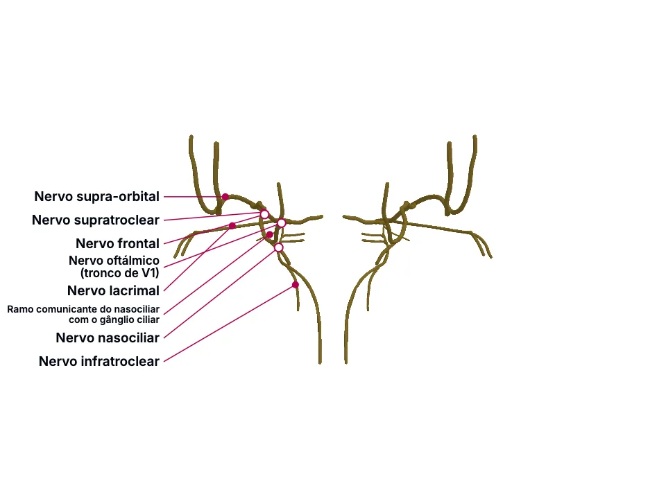
O nervo oftálmico (V1), menor divisão do trigêmeo, se ramifica em lacrimal, frontal e nasociliar - este último ligando pele do nariz e córnea (sinal de Hutchinson) e convergindo, na fissura orbital superior, com os nervos que movem o olho.
Escopo e método deste resumo
O nervo oftálmico (V1), menor divisão do trigêmeo, se ramifica em lacrimal, frontal e nasociliar - este último ligando pele do nariz e córnea (sinal de Hutchinson) e convergindo, na fissura orbital superior, com os nervos que movem o olho.
Material educacional escrito com apoio de IA e conferido por revisão automatizada, sem revisão médica nomeada. Não substitui o livro-texto nem orientação clínica.
Essencial
O nervo oftálmico (V1) é a menor das três divisões do trigêmeo: 'The ophthalmic nerve is the smallest trigeminal nerve division.' Ele sai da fossa craniana média pela fissura orbital superior e, no ápice orbitário, se divide em três ramos nomeados: 'The nerve then exits the skull through the superior orbital fissure and divides in the orbital apex into the lacrimal, frontal, and nasociliary nerves.'
Essa divisão em três ramos - lacrimal, frontal e nasociliar - não vem de uma única fonte: um capítulo independente, sobre herpes zoster oftálmico, descreve exatamente a mesma lista: 'V1 is subdivided into 3 branches: the frontal nerve branch, the nasociliary nerve branch, and the lacrimal nerve branch.' E um terceiro capítulo, sobre a anatomia do nariz, repete a mesma lista de três: 'The ophthalmic division has three main branches: the lacrimal, frontal, and nasociliary.' Três fontes StatPearls diferentes concordando na MESMA lista específica de três nomes - não apenas no mesmo assunto geral.
O nasociliar é um dos três ramos terminais de V1, mas segue um trajeto diferente dos outros dois dentro da fissura orbital superior: passa DENTRO do anel tendíneo comum, junto dos nervos oculomotor e abducente: 'Passing through the superior orbital fissure and the common tendinous ring are the superior and inferior divisions of CN III (oculomotor), the nasociliary branch of CN V1, sympathetic roots of the ciliary ganglion, and CN VI (abducens).' - enquanto lacrimal e frontal passam FORA desse anel, junto do troclear: 'Passing through the superior orbital fissure into the orbit and above the common tendinous ring are the lacrimal nerve of CN V1, the frontal nerve of CN V1, CN IV (trochlear nerve), and the superior ophthalmic vein.'
O nasociliar é o único ramo de V1 que inerva, ao mesmo tempo, a ponta do nariz e a córnea: 'The nasociliary branch innervates the tip of the nose, cornea, and other ocular structures.' É essa dupla inervação - MESMO nervo, dois territórios - que dá valor preditivo a uma erupção na pele do nariz: 'The Hutchinson sign—vesicles on the tip or side of the nose—indicates nasociliary nerve involvement and correlates with a higher likelihood of ocular complications.'
O nasociliar - um dos três ramos terminais do nervo oftálmico (V1) - dá origem a ramos mais finos, cada um com seu próprio forame ósseo: 'The anterior ethmoidal foramen is in the frontoethmoidal suture, while the posterior ethmoidal foramen is located at the junction of the orbit's roof and medial wall. These two foramina transmit the anterior ethmoidal vessels and nerve and the posterior ethmoidal vessels and nerve, respectively.' O etmoidal anterior leva sensibilidade à porção anterossuperior do nariz interno e à mucosa septal anterior: 'The anterior ethmoidal nerve (branch of the nasociliary) provides sensation to the anterosuperior aspect of the internal nose and the anterior nasal septal mucosa.'
O nasociliar também dá um ramo comunicante ao gânglio ciliar - descrito, num capítulo dedicado à anatomia desse gânglio, sob outro nome: 'Here it is embedded in orbital fat and is connected to the nasociliary nerve.' O gânglio tem três raízes distintas: 'Three roots of the ciliary ganglion are attached to its posterior border. These roots are motor root, sensory root, and sympathetic root.' e a raiz SENSITIVA - a mesma estrutura que o bundle 3D chama de 'ramo comunicante do nasociliar com o gânglio ciliar' - deriva exatamente do nasociliar: 'The sensory root derives from the nasociliary nerve. Sensory fibers from eyeball pass through ganglion uninterrupted. It carries general sensations from the cornea, iris, and ciliary body.'
O frontal e o nasociliar são dois dos três ramos terminais do nervo oftálmico (V1). O supra-orbital é ramo sensitivo do FRONTAL: 'The supraorbital nerve is a sensory branch of the frontal nerve, which arises from the ophthalmic division (V 1 ) of the trigeminal nerve.' Depois de emergir, sobe para inervar testa, pálpebra superior e couro cabeludo anterior. O supratroclear também é ramo do frontal (árvore confirmada contra referência de anatomia geral, ver nota acima) e cobre uma porção menor e mais medial da testa: 'After emerging, it ascends to supply cutaneous sensation to the forehead, upper eyelid, and anterior scalp, whereas the supratrochlear nerve supplies a smaller and more medial portion of the forehead.' Já o infratroclear NÃO nasce do frontal - nasce do nasociliar: 'The infratrochlear nerve arises from the nasociliary nerve and provides innervation of the superior aspect of the external nose.'
Apesar dessa origem diferente, os três são agrupados aqui pelo mesmo motivo clínico: juntos, marcam a fronteira sensitiva da testa, pálpebra superior e raiz do nariz usada em bloqueio anestésico local: 'Clinicians commonly perform this block for procedures involving the forehead and periorbital soft tissues, and the technique has also been used to treat selected headache disorders and ophthalmic herpetic neuralgia.'
Na clínica
Cada um dos três ramos pode ser acometido separadamente - sinal de que são unidades anatômicas distintas, não um bloco único: 'Any or all of these nerve branches may be affected by HZO' No mesmo sentido, V1 como um todo é primariamente sensitivo, cobrindo testa, olho, nariz e couro cabeludo: 'CN V1 is primarily a sensory nerve supplying the scalp, eye, nose, and forehead.' - detalhamento confirmado por um QUARTO capítulo independente, sobre os ossos da órbita: 'The ophthalmic nerve is the first division of the trigeminal nerve (CN V) and is responsible for conveying sensory information from the globe, upper eyelid, lacrimal gland, conjunctiva, nasal mucosa, frontal sinus, external nose, and scalp.'
Por isso, vesículas na ponta ou lateral do nariz (o sinal de Hutchinson) indicam envolvimento do nasociliar e maior risco de complicação ocular: 'The presence of the Hutchinson sign indicates nasociliary branch involvement of V1, which confers a higher risk for ocular involvement.' Mas o sinal não é uma condição necessária - sua AUSÊNCIA não descarta doença ocular: 'The Hutchinson sign (vesicles on the nose tip) strongly predicts ocular involvement, but its absence does not exclude eye disease.'
Essa raiz sensitiva é o que permite que uma lesão do nasociliar (ou do próprio ramo comunicante) altere a sensibilidade da córnea, da íris e do corpo ciliar - estruturas do olho, enquanto o etmoidal anterior leva a sensibilidade ao nariz, mostrando como um único ramo (o nasociliar) distribui sensibilidade para estruturas anatomicamente distantes entre si (olho e nariz) através de seus sub-ramos.
Um bloqueio de supraorbital e supratroclear anestesia toda a testa e a pálpebra superior para debridamento, sutura de laceração complexa ou alívio de dor aguda de herpes zoster, sem distorcer os planos de tecido como a infiltração local faria: 'After emerging, it ascends to supply cutaneous sensation to the forehead, upper eyelid, and anterior scalp, whereas the supratrochlear nerve supplies a smaller and more medial portion of the forehead.' - 'Clinicians commonly perform this block for procedures involving the forehead and periorbital soft tissues, and the technique has also been used to treat selected headache disorders and ophthalmic herpetic neuralgia.'
Abrir o aprofundamento
Aprofundamento
A fissura orbital superior é a via de saída comum para múltiplos nervos entre o crânio e a órbita: 'The middle portion of the superior orbital fissure contains the cranial nerves III, IV, and VI and the ophthalmic division of the trigeminal nerve (V1).' Quando essa região é acometida por massa, trauma ou inflamação, o quadro resultante - a síndrome do ápice orbitário - envolve vários pares cranianos ao mesmo tempo: 'The orbital apex syndrome is a complex multiple cranial nerve pathology that results from the involvement of the bony orbital apex.'
Nervo oftálmico (V1) e seus ramos - contribuem o componente SENSITIVO do quadro: dor e parestesia na testa e na pálpebra superior. Nenhum ramo de V1 move o olho; ele só sente a região ao redor dele.; Nervos oculomotor, troclear e abducente - contribuem o componente MOTOR: restrição do movimento ocular (os três juntos movem o globo em quase todas as direções), e o oculomotor especificamente também carrega fibras pupilares - daí a anisocoria. Juntos com V1, esses três nervos formam o quadro misto sensitivo+motor característico da síndrome: 'The clinical signs of orbital apex syndrome are due to the involvement of the optic nerve, oculomotor nerve, trochlear nerve, abducens nerve, and the ophthalmic branch of the trigeminal nerve.' - 'restricted ocular movement due to the involvement of the oculomotor, trochlear, and abducens nerve, facial pain and paresthesia over the forehead and the upper lid due to the involvement of the ophthalmic division of trigeminal nerve, and anisocoria due to the involvement of the pupillary fibers.'; A síndrome da fissura orbital superior - o mesmo grupo de nervos (III, IV, VI e V1), mas SEM envolvimento do nervo óptico - se diferencia da síndrome do ápice orbitário por esse único detalhe, a poupança da visão: 'is characterized by the involvement of the III, IV, VI, and the ophthalmic division of the trigeminal nerve. Still, it differs from OAS with the sparing of the optic nerve.'
Teste-se antes de seguir
Uma paciente perde a sensibilidade de toda a testa, da pálpebra superior e do dorso do nariz de um lado, sem nenhuma alteração de movimento ocular. Qual nervo explica esse padrão, e em quantos ramos terminais nomeados ele se divide no ápice orbitário?
A resposta fica na Avaliação, sem spoiler aqui.
Responder na AvaliaçãoFontes (7)
- Huff T, Weisbrod LJ, Daly DT. Neuroanatomy, Cranial Nerve 5 (Trigeminal). [Updated 2024 Apr 20]. In: StatPearls [Internet]. Treasure Island (FL): StatPearls Publishing; 2026 Jan-.
The ophthalmic nerve is the smallest trigeminal nerve division.
- Minor M, Gurnani B, Payne E. Herpes Zoster Ophthalmicus. [Updated 2025 Sep 14]. In: StatPearls [Internet]. Treasure Island (FL): StatPearls Publishing; 2026 Jan-.
V1 is subdivided into 3 branches: the frontal nerve branch, the nasociliary nerve branch, and the lacrimal nerve branch.
- Galarza-Paez L, Marston G, Downs BW. Anatomy, Head and Neck, Nose. [Updated 2023 Jul 24]. In: StatPearls [Internet]. Treasure Island (FL): StatPearls Publishing; 2026 Jan-.
The ophthalmic division has three main branches: the lacrimal, frontal, and nasociliary.
- Shumway CL, Motlagh M, Wade M. Anatomy, Head and Neck, Orbit Bones. [Updated 2023 Jul 24]. In: StatPearls [Internet]. Treasure Island (FL): StatPearls Publishing; 2026 Jan-.
The ophthalmic nerve is the first division of the trigeminal nerve (CN V) and is responsible for conveying sensory information from the globe, upper eyelid, lacrimal gland, conjunctiva, nasal mucosa, frontal sinus, external nose, and scalp.
- Bhardwaj N, Joshi A. Neuroanatomy, Ciliary Ganglion. [Updated 2023 Jul 24]. In: StatPearls [Internet]. Treasure Island (FL): StatPearls Publishing; 2026 Jan-.
Here it is embedded in orbital fat and is connected to the nasociliary nerve.
- Napier A, Dua A, Taylor A. Supraorbital Nerve Block. [Updated 2026 Jul 5]. In: StatPearls [Internet]. Treasure Island (FL): StatPearls Publishing; 2026 Jan-.
The supraorbital nerve is a sensory branch of the frontal nerve, which arises from the ophthalmic division (V 1 ) of the trigeminal nerve.
- Mohankumar A, Gurnani B. Orbital Apex Syndrome. [Updated 2023 Jun 11]. In: StatPearls [Internet]. Treasure Island (FL): StatPearls Publishing; 2026 Jan-.
The middle portion of the superior orbital fissure contains the cranial nerves III, IV, and VI and the ophthalmic division of the trigeminal nerve (V1).
Onde isso cai na prática
1 caso clínico do acervo exige este tema. Veja onde ele decide a conduta.
Dor em queimação e vesículas na hemiface direita em uma mulher de 71 anos na UBS
Dermatologia
Abrir o caso grátis
Mais resumos de Anatomia
Depois de ler, use o que aprendeu
Atlas
Veja as estruturas do tema em 3D e navegue por camadas.
Caso clínico
Aplique o conteúdo numa decisão de plantão.
Exame
Interprete o resultado que fecha o raciocínio.
Avaliação
Responda as perguntas dos blocos e veja onde você está.
Tutor de IA
Tire a dúvida que o texto não resolveu.
Comece grátis: crie a conta e prove uma amostra do Atlas em 3D, do caso clínico, do exame, da avaliação e do tutor de IA. Sem cartão de crédito.
Começar grátis